Cellular science · Science
The power of the mitochondria: how red light recharges your cells
Mitochondria are not just the textbook powerhouse of the cell. They are the decision-makers of your biology — and they happen to be light-sensitive.
18 August 2026 · 14 min read
Key takeaways
- Mitochondria convert food and oxygen into ATP, the universal energy currency of every human cell.
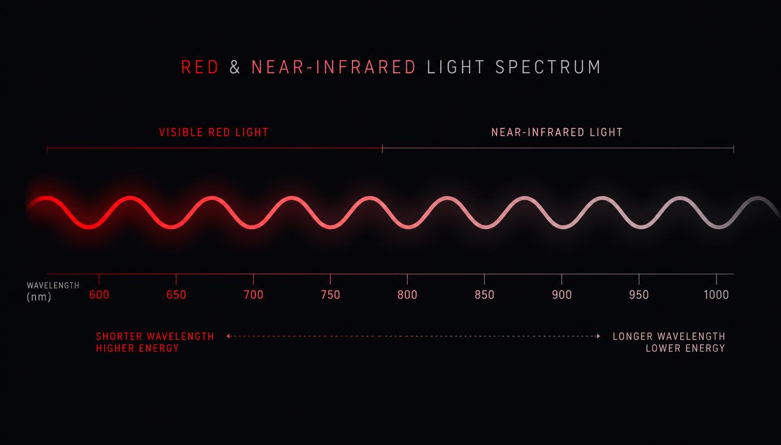
- Red (630–660 nm) and near-infrared (810–850 nm) light are absorbed by cytochrome c oxidase, the fourth complex of the respiratory chain.
- That absorption displaces inhibitory nitric oxide, restores electron flow and increases ATP output.
- A short burst of signalling reactive oxygen species triggers repair genes rather than damage.
- Consistency beats intensity: 10–20 minutes, most days, at a sane dose.
What mitochondria actually do
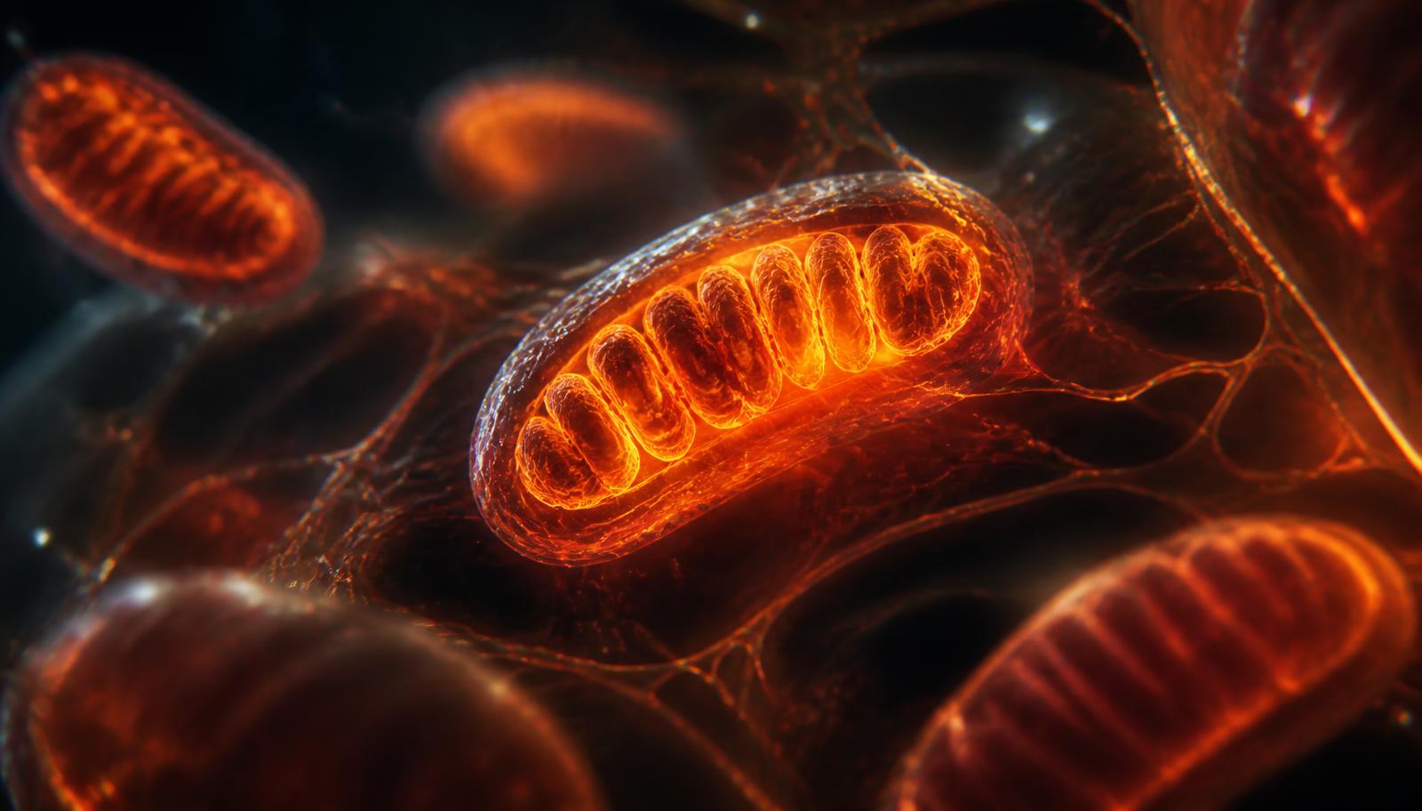
Roughly two billion years ago, a bacterium was engulfed by another cell and, instead of being digested, stayed. That ancient bargain became the mitochondrion — an organelle with its own circular DNA, its own membranes and its own agenda. Today a single muscle or heart cell can host thousands of them, occupying up to 40% of its internal volume.
Their headline job is oxidative phosphorylation: taking electrons stripped from the food you eat, passing them down a chain of four protein complexes embedded in the inner mitochondrial membrane, and using the energy released to pump protons across that membrane. The resulting electrochemical gradient drives ATP synthase like water drives a turbine. The output is adenosine triphosphate — ATP — the molecule that pays for muscle contraction, nerve signalling, protein synthesis, DNA repair and collagen production.
~50 kg
ATP recycled per day in an average adult
1000s
Mitochondria per high-demand cell
90%
Of cellular oxygen consumed by mitochondria
But energy is only half the story. Mitochondria also regulate calcium buffering, steroid hormone synthesis, innate immune signalling, and apoptosis — programmed cell death. When mitochondrial performance drops, cells do not simply slow down: they change behaviour. Fibroblasts make less collagen. Neurons fire less efficiently. Muscle recovers more slowly. Skin looks tired because it literally is.
Why mitochondrial performance declines
Mitochondrial function is not fixed. It is a moving target shaped by age, sleep, movement, inflammation, medication and light exposure. Three mechanisms dominate the decline.
1. Nitric oxide competition
Under stress, hypoxia or chronic inflammation, nitric oxide accumulates and binds to cytochrome c oxidase — the final complex of the electron transport chain — at the same site oxygen should occupy. Electron flow slows. Proton pumping slows. ATP output falls even though fuel and oxygen are available.
2. Oxidative damage accumulation
Electron leak produces reactive oxygen species. In small, pulsed amounts these are useful signals. Sustained at high levels they oxidise mitochondrial lipids, proteins and the mitochondrial DNA sitting a few nanometres from the source, with no protective histones and limited repair machinery.
3. Loss of mitochondrial quality control
Healthy tissue constantly recycles damaged mitochondria (mitophagy) and builds new ones (biogenesis, largely via PGC-1α). With age and sedentary living, both processes slow, so damaged organelles linger and the average quality of the mitochondrial pool falls.
The practical consequence
Low cellular energy rarely feels like a diagnosis. It feels like slower recovery, duller skin, foggier afternoons and worse sleep — the everyday symptoms of a metabolism running with the handbrake half on.
How light physically reaches your mitochondria
Skin is not opaque. It is a scattering, partially transparent medium, and its transparency depends heavily on wavelength. Below about 600 nm, haemoglobin and melanin absorb light aggressively, so blue and green photons stop in the epidermis. Above about 1100 nm, water absorption takes over and photons are converted mostly to heat.
Between those limits sits the optical window — roughly 600 to 1100 nm — where absorption by the main chromophores is at its lowest and photons can travel millimetres to centimetres into tissue. Red light around 630–660 nm reaches the dermis, where fibroblasts build collagen. Near-infrared light at 810–850 nm penetrates further, reaching muscle, joints, deeper vasculature and, with sufficient irradiance, the cortical surface of the brain.
| Wavelength | Typical depth | Primary target |
|---|---|---|
| 630–660 nm (red) | 1–5 mm | Epidermis, dermal fibroblasts, capillaries |
| 810–830 nm (NIR) | 10–30 mm | Muscle, joints, neural tissue |
| 840–860 nm (NIR) | 20–40 mm | Deep muscle, connective tissue |
This is why serious devices combine both bands. Red alone treats the surface. Near-infrared alone skips it. Together they cover the full depth gradient of a treated area, which is exactly what a systemic effect requires.
Cytochrome c oxidase: the photoreceptor you didn't know you had
For light to do anything biological, a molecule has to absorb it. In photobiomodulation the primary acceptor is cytochrome c oxidase (CCO), complex IV of the respiratory chain. CCO contains two copper centres and two haem groups whose absorption peaks land almost exactly on the wavelengths used in red light therapy — a striking coincidence that explains why the field converged on 660 nm and 850 nm.
- A photon in the red or near-infrared band is absorbed by a copper centre of cytochrome c oxidase.
- The absorbed energy weakens the bond holding inhibitory nitric oxide at the enzyme's active site.
- Nitric oxide is released into the cytosol, where it acts as a local vasodilator, improving microcirculation.
- Oxygen regains access to the catalytic site; electron transport accelerates.
- The proton gradient across the inner membrane steepens and ATP synthase output rises.
- A brief, controlled rise in reactive oxygen species acts as a redox signal, not as damage.
That last step matters more than most marketing admits. The transient ROS pulse activates redox-sensitive transcription factors — NF-κB, Nrf2, AP-1 — which upregulate antioxidant enzymes, heat shock proteins and growth factors. The net effect over days is a cell that is better defended than before the exposure. This is hormesis: a small, well-dosed stressor producing a net adaptive gain.
Photobiomodulation does not add energy to the body. It removes a brake from a system that was already capable of producing more.
What changes downstream, tissue by tissue
A rise in ATP availability is invisible on its own. What you notice are the downstream expenditures your cells can suddenly afford.
Skin
Dermal fibroblasts are ATP-hungry. Collagen I and III synthesis, elastin assembly and the balance of matrix metalloproteinases all improve when energy is not the limiting factor. Clinically this shows up as firmer texture, more even tone and better hydration over 8–12 weeks of consistent use.
Muscle and connective tissue
Near-infrared exposure before or after training is associated with reduced markers of muscle damage such as creatine kinase, less delayed-onset soreness and faster restoration of peak force. The mechanism is a combination of improved mitochondrial output, better microcirculation via released nitric oxide, and modulated inflammatory signalling.
Nervous system and sleep
Transcranial and evening whole-body sessions have been studied for their effect on subjective sleep quality and next-day alertness. The plausible pathway runs through improved cerebral blood flow, mitochondrial support in high-demand neurons and — importantly — the absence of the short-wavelength blue light that suppresses melatonin.
Circulation
Nitric oxide released from CCO diffuses into surrounding smooth muscle and relaxes it. Local perfusion rises, delivering more oxygen and nutrients and clearing metabolic waste faster. This is the quiet, underrated half of the red light story.
Dose: the part almost everyone gets wrong
Photobiomodulation follows a biphasic dose response, often described as the Arndt-Schulz curve. Too little light produces no measurable effect. The right dose produces the full effect. Substantially more than the right dose produces less effect than the right dose — not damage in most cases, but a wasted session.
Dose is energy density, measured in joules per square centimetre: irradiance (mW/cm²) multiplied by time (seconds), divided by 1000. Most clinical protocols for skin and superficial tissue land between 3 and 10 J/cm². Deeper musculoskeletal targets often use 10 to 60 J/cm² at the surface, because scattering and absorption reduce what actually arrives at depth.
| Goal | Distance | Session | Frequency |
|---|---|---|---|
| Skin, tone and texture | 20–30 cm | 10 min | 5–7× per week |
| Muscle recovery | 15–20 cm | 10–15 min | After training |
| Evening wind-down | 30–40 cm | 10–20 min | Nightly, 1–2 h before bed |
More is not better
Doubling your session length rarely doubles the benefit and can flatten it. Treat light like training volume: consistent, moderate and repeated beats occasional and extreme.
Turning the science into a daily ritual
The biology is only useful if the behaviour is sustainable. The people who see the clearest results are not the ones with the most powerful device — they are the ones who attach the session to something they already do every day.
- Anchor the session to an existing habit: after the shower, before brushing teeth, during the last podcast episode of the day.
- Keep the distance and duration constant so you are comparing like with like over weeks.
- Expose bare skin. Fabric scatters and absorbs a surprising amount of red light.
- Photograph the same area in the same light every two weeks; perception drifts, images do not.
- Give it 8 to 12 weeks before judging. Collagen remodelling runs on a slower clock than motivation.
Mitochondria respond to patterns, not to single events. Sleep, movement, cold, heat, fasting and light are all inputs into the same adaptive system. Red light is one lever among several — but it is the one you can pull for ten quiet minutes without changing anything else about your day.
Frequently asked questions
How does red light therapy affect mitochondria?
Red and near-infrared photons are absorbed by cytochrome c oxidase, the fourth complex of the mitochondrial electron transport chain. The absorbed energy releases inhibitory nitric oxide from the enzyme, restoring oxygen binding and electron flow. The result is a steeper proton gradient and increased ATP production, plus a brief signalling burst of reactive oxygen species that activates cellular repair pathways.
Which wavelengths are best for mitochondrial support?
The best-studied bands are 630–660 nm in the visible red range and 810–850 nm in the near-infrared. Red penetrates a few millimetres and targets skin and capillaries; near-infrared reaches muscle, joints and deeper tissue. Combining both covers the full depth gradient.
How long before I notice anything?
Acute effects like warmth, local circulation and post-session calm are immediate. Recovery benefits typically appear within one to two weeks. Structural changes such as skin firmness and tone follow collagen turnover and generally need 8 to 12 weeks of consistent use.
Can you overdo red light therapy?
Yes, in the sense that the dose response is biphasic: beyond an optimal energy density the benefit plateaus and then diminishes. Longer sessions are not better sessions. Follow the recommended distance and duration rather than maximising exposure.